
Compiled by I. Abdullah.
This post is “work in progress”. Last updated on the 28th April 2013, 21:00 Any comments are most welcome.

The Qur’an mentions that human development passes through a number of distinct stages (Qur’an 39:6 and 71:14). These stages are descriptive of the embryo’s external appearance and have been assigned the following names as we read in Surah Al-Mu’minoon (The Believers) 23: 12-14:
And We (God) created man from a quintessence (gentle extraction) of clay. We then placed him as a nutfah (drop) in a place of settlement, firmly fixed (i.e. the womb). Then We made the drop into an ‘alaqah (clinging form), and then We changed the clinging form into a mudghah (chewed-like form), then We made out of that chewed-like form, izam (skeleton, bones), then We clothed the bones with lahm (muscles, flesh), then We (ansha’ nahu), caused him to grow and come into being and attain the definitive (human) form. Blessed be God, the Perfect creator.
This article briefly describes the Izam (skeleton, bones) and Lahm (muscles, flesh) stages of development.
The Izam stage

then We made out of that chewed-like form, izam (skeleton, bones)
The stage of development after the chewed-like form (mudghah) is referred to as izam which means “bones” (3) and the fetus does indeed acquire a cartilaginous skeleton of bones. In the 6th week the cartilaginous skeleton begins to form and the embryo acquires a soft skeleton as we see in Figure 2.
“Formation of bone does not begin uniformly throughout the body. Rather, there is a sequential appearance of bony tissue. However, in the 7th week the spreading development of the skeleton occurs. Bone development in the limbs commences in the limb buds from mesochymal cells. Primary ossification centers appear in the femur during week 7 and in the sternum (breast bone) and the maxilla (upper jaw) in weeks 8-9.” (4)
Mesenchymal bones are made out of connective tissue which become cartilaginous, and then they become ossified and become (solid) bones. Mesenchymal bones form during the fifth week as condensations of mesenchyme appear in the limb buds (Figure 3 A to C). During the sixth week, the mesenchymal bone models in the limbs undergo chondrification to form hyaline cartilage bone models as wee see in Figure 3 D and E.

The timing of the izam phase has been mentioned in the following Hadith (Sahih Muslim, Kitab Al-Qadar):

When 42 nights have passed from the time of the nutfah (time of conception), God sends an angel to it, who shapes it and makes its ears, eyes, skin, muscles and bones…
“In the early part of this (izam) phase, the embryo takes on a human appearance (tasweer adami), and the hadith describes this with the word “shapes”. Before the 42nd day, it is difficult to distinguish the human embryo from the embryos of many animals, but at this time it becomes clearly distinguishable in its appearance.” (4)
The formation of the skeleton gives the embryo its human shape.
The Lahm stage
then We clothed the bones with lahm (muscles, flesh)…
The next stage is the lahm stage (muscles, flesh) stage. The lahm stage is characterized by clothing the bones with muscles (al-kisa’ billahmn). During the lahm stage, the embryo develops human features, and the various organs assume their proper positions and are better proportioned. Thus during this stage the effect on the embryo of muscles clothing the bones is manifest in its external appearance.
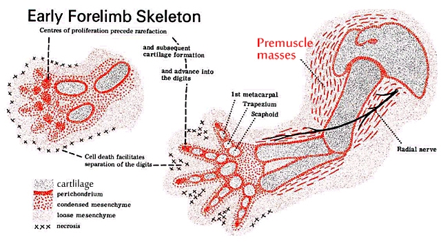
“Soon after the cartilaginous models of the bones have been established, the myogenic cells, which have now become myoblasts, aggregate to form muscle masses on the ventral [front] and dorsal [back or posterior] aspects of the limbs.” (5)
Profs. Smith and Williams state that:
“Muscles are first indicated as premuscle masses of condensed mesenchyme in the base of the limb bud [Figure 4]. Myoblasts become spindle-shaped and arrange in parallel bundles in which they fuse end to end, forming long multinucleate myotubes. Myofibrils appear within the myotubes and, increasing in number and size, develop cross-straitions.The final number of fibres in a muscle is reached sometime before birth. Muscles then grow by the increase in length and thickness of individual fibres and by the addition of myoblasts at their ends. As differentiation proceeds proximodistally [from the shoulder to the tips of the digits] within the limb bud premuscle masses are formed dorsal and ventral to the developing bone. Flextor and adductor muscles develop in the ventral mass and extensor and abductor muscles in the dorsal mass.” (6)
The primordia from which the bones and muscles develop are formed before the 7th week, differentiation of the skeleton occurs in the 7th week and differentiation of the muscles occurs next in the 8th week:
“Although precursor cells (myoblasts, or primitive muscle cells) are present adjacent to developing bone, differentiation into skeletal muscle attachments occur after the ossification process in the shaft and ends of the bones has begun.
A major developmental landmark during the eighth week is the lahm stage, which describes the myogenesis (muscle formation) period, and which marks the development of definitive muscles in the trunk and limbs and the beginning of movement. The muscles take their position around the bones (“clothing the bones”) and continue the process of straightening and smoothing (taswiyah) which began in the izam stage.” (4)
“[The] muscles and tendons become attached to the bony structures so that they can produce their actions across the joints.” (5)

The Lahm stage begins from the about the end of the 7th week to the end of the 8th week and comes immediately after the izam stage.
Figure 2 and Figure 5 show a human embryo at Stage 20 (about days 50 – 51 days) showing the cartilage skeleton and skeletal musculature.(2) For further information on bone and muscle development see Embryology in the Qur’an: Bone and Muscle Development (4).
Note From a Reader: The development of the Meckel’s cartilage
The abstract of a paper published in Anatomical Science International on development of the Meckel’s cartilage may be of interest to your readers. The Meckel’s cartilage is a cartilaginous bar in the fetus around which the mandible (jaw bone) develops. Part of Meckel’s cartilage develops into the malleus (an ear ossicle) in the adult. Note the sequence of the development of the Meckel’s cartilage:
“The Meckel’s cartilage itself and the mandible are derived from the first branchial arch, and their development depends upon the contribution of the cranial neural crest cells. The prenatal development of the Meckel’s cartilage, along with its relationship to the developing mandible and the related structures, were studied histologically in human embryos and fetuses. The material was obtained from a collection of the Department of Anatomy, and laboratory procedures were used to prepare sections, which were stained according to standard light-microscopy methods. The formation of the Meckel’s cartilage and its related structures was observed and documented. Some critical moments in the development of the Meckel’s cartilage are suggested. The sequential development of the Meckel’s cartilage started as early as stage 13 (32 days) with the appearance of condensation of mesenchymal cells within the mandibular prominence. During stage 17 (41 days), the primary ossification center of the mandible appeared on the inferior margin of the Meckel’s cartilage. The muscular attachments to the Meckel’s cartilage in embryos were observed at stage 18 (44 days).“
Wyganowska-Świątkowska M, Przystańska A. The Meckel’s cartilage in human embryonic and early fetal periods. Anat Sci Int. 2011 Jun;86(2):98-107. doi:10.1007/s12565-010-0093-3. Epub 2010 Aug 27. PubMed PMID: 20799009.
RELATED VIDEOS & ARTICLES
- Video: Embryology in the Qur’an – Bone and Muscle development with Dr. Keith L. Moore
- Video: Embryology in the Qur’an – Bone and Muscle development with Dr. TVN Persaud
- Human Development in the first 40 days
- Human Development after the 42nd day
Footnotes
(1) The Multi-Dimensional Human Embryo, Stage 20, Day 50.
(2) Ulrich Drews, Color Atlas of Embryology, Thieme Medical Publishers, 1995. Page 98-99. Showing the cartilaginous skeleton of an embryo at stage 20 (about day 50 – 51, 18 – 22 mm).
(3) Izam meaning “skeleton, bones”. The term encompasses both cartilage and ossified (hard) bone. This post refers to the development of the appendicular skeleton which consists of the pectoral and pelvic girdles and the limb bones.
(4) G. C. Goeringer, A. A. Zindani, M. A. Ahmed, Embryology in the Qur’an: Bone and Muscle Development. https://islam-papers.com/2012/03/06/bone-and-muscle/
(5) John Allan and Beverley Kramer, The Fundamentals of Human Embryology. 2nd Edition. Wits University Press. 2010, page 148.
(6) C.P. Wendell Smith and P.L. Williams, Basic Human Embryology, 3rd Edition, London, Pitman Publishing Ltd., 1984, p. 114.
(7) Video: Embryology in the Qur’an by Dr. Keith Moore (1990). University of Illinois, Chicago, USA. https://islam-papers.com/2012/03/07/moore1/

“Soon after the cartilaginous models of the bones have been established, the myogenic cells, which have now become myoblasts, aggregate to form muscle masses on the ventral and dorsal aspects of the limbs.”
Right; so how could PZ Myers simplistically claim that the two processes occur simultaneously and, worse still, how could Adnan Rashid have agreed by claiming that the Qur’an supports both processes occurring sequentially and simultaneously?!
To the contrary, PZ Myers et al., must contradict the Qur’an even to the point of denying the evidences. If a Muslim was to point out to sunbathing militant atheists like Myers that the sun is shining down in the bright blue sky, they’d deny that too just to contradict the Muslim!
Some people have misunderstood what Moore has written and lectured about. From my limited understanding, the skeleton first appears as cartilage bone models. See http://islampapers.com/2012/04/01/bone-and-muscle-2/ and view the illustrations.
Muscles then attach to bony structures (ossified bone) – which means that some ossified bone must be present for the muscles to attach to? When Moore talks about bone, some misunderstand and only think of hard (ossified) bone. “Bone” can also be soft (cartilage) bone too. Even in the English language we can talk of fish bone and chicken bone.
“[W]hich means that some ossified bone must be present for the muscles to attach to”
Well, that makes logical sense to me.
Mash’allah very impressed with your work brothers, may Allah reward you with good.
PZ Myers is correct that the processes occur in parallel. Formation of cartilage models, muscle and bone happen over an extended, overlapping period. Both the start of chondrification and grouping of myoblasts occur in a proximal-distal order in the limb.
Yes, the myoblasts aggregate into 2 distinct groups as chondrification (differentiation into cartilage of the condensed mesenchymal core in the limb) is beginning, but these cells that fuse to become muscle had already migrated into the limb (day 26) and around the condensing mesynchyme in a proximal-distal order before chondrification begins in the upper part of the limb (day 37) and lower part of the limb (day 41). You need to read a more detailed account to see this than the above quotes. See Galjaard, R.J.H 2003. Mapping Studies of Congenital Limb Anomalies. Page 16 (can be read as a pdf at http://repub.eur.nl/res/pub/10474/030924_Galjaard,%20Robert-Jan%20Harmen.pdf). Read carefully, this more detailed account doesn’t contradict the quotes in the above article. Note also that merely the start of chondrification does not mean that the cartilage models have yet formed such that you could reasonably say bones have formed.
This diagram (strictly speaking for mice, but considered a good analog for humans in this respect) helps us appreciate the complexity of the process:
http://php.med.unsw.edu.au/embryology/index.php?title=Musculoskeletal_System_-_Muscle_Development#Mouse_Limb_Muscle
All this is in contrast to an Muslim debater who a year ago wrote a paper (after an encounter with PZ Myers) that took certain ambiguous quotes to incorrectly claim that the precursor muscle cell migration (not aggregation) along the limb happens after the cartilage models have formed so that he could vaguely claim it to be a process of “clothing” after bones have formed.
But Somitic, you’ve missed the forest for the trees. The reason why I said P.Z. Myers claim was “simplistic”, and not untrue in the absolute sense, was because of the overall import of his assertion, which, of course, was to contradict the claim made in the Qur’an that the bones were formed first in order for the skin to clothe them. Unlike what some of his fellows have acknowledged in their more honest and nuanced evaluation of this subject (as cited in the above article), P.Z. Myers decided to adopt an absolutist position in this regard. His blanket claim gives the impression that the entire process occurs simultaneously when it clearly does not; there was more to this than meets the eye, and as an expert who presumably knows his field he should have known this and, therefore, should not have made such a simplistic assertion. I would hazard a guess and say that the reason why he made this rigid and inflexible remark was probably because of the modus operandi of most militant atheists of wanting to contradict the Qur’an and seek to defeat the Muslims hook or by crook to the point of absurdity.
This is a miracle of Science and Quran, but the hate-mongers of Islam wouldn’t
understand it. How much more proof and evidence they need to prove that there is
One GOD, Allah-Creator and Quran is HIS last divine Revelation.
Salaam
Sis. G.
Thanks for this website! MashAllah! Salam
Pingback: Islam and science: The road to renewal - ECONOMIST - Page 11
Pingback: Naswar Corner - Page 1824
This is a clear PROOF that the QUR’AN is from The CREATOR of Man!
ALLAH HU AKBAR KABIRA
Reblogged this on Perjalanan Hati and commented:
Very interesting paper, especially to the musculoskeletal topic which I teach.
Assalamualaikum,What about the hadith in sahih bukhari 3208 and muslim 2643 which states that for every stage is 40 days timeline for each stage, that’s incorrect cuz the hadith states from a nutfah take a 40 days in mother womb which is incorrect cuz it takes 6 days approximately, and it says that alaqah(leech like form and clinging) take 40 days too which is incorrect cuz the embryo at day 40 is more like a bean and the leech like form end in day 20 , and for the muduqah(chewed like form) 40 days too which is now day 80(if we added them up) we can say which is incorrect cuz the embryo doesn’t look like a chewed like form in day 80 ,and also the embryo heartbeat(wanafakh fih alruwh) at day 120 or 160(if we added them all) which is incorrect cuz embryo heartbeat at week 6-8
Jzk for your effort , waiting for your replay.
See Human Development in the first 40 days | The Islam Papers