
This is an early paper by Dr. Keith L. Moore published in 1982. Some of his interpretations have evolved since then. See his later works: A Scientist’s Interpretation of References to Embryology in the Qur’an (1986) and Dr. Keith Moore at the University of Illinois, Chicago, USA (1990)
The paper is from Arabization and Medical Education, pp. 51-58. Proceedings from the Seventh Saudi Medical Conference, King Faisal University, May 3-6 1982.

Keith L. Moore, Ph.D., F.I.A.C.
Professor of Anatomy and Chairman of the Department, Faculty of Medicine, University of Toronto, Toronto, Ontario, Canada.
Human beings have always been interested in where they came from and how they developed before birth. We know from the earliest records that primitive peoples realized that the birth of a baby was the sequel to sexual union or intercourse. However, for many centuries the idea about human prenatal development were based on speculation and mysticism. The absence of knowledge about embryological processes and the dominating influence of superstition resulted in a non-scientific approach to human development.
As far was we know, Aristotle wrote the first embryology book in the 4th century BC. In it he recorded some observations on comparative embryology, especially on the general progress of the developing chick. He promoted, however, the incorrect idea that the human embryo developed from a formless mass that resulted from the union of the semen with the menstrual blood.
Scientific knowledge of embryology did not progress significantly for nearly 2000 years. It was not until the close of the 17th century, when the microscope was developed, that the early stages of human development could be effectively studied. After it was possible to examine cells under the microscope, it was reasoned in the 18th century that development resulted from the growth and differentiation of embryonic cells.
Almost a year ago I was consulted about the meaning of certain verses in the Qur’an and some sayings in the Hadiths which referred to human reproduction and embryological development.
I was amazed at the scientific accuracy of these statements which were made in the 7th century AD. I have selected verses and sayings for which I shall provide personal interpretations based on my knowledge of embryological history and of the modern science of embryology.
The realization that the embryo develops in stages in the uterus (figure 1) was not discussed or illustrated until the 15th century AD., although Galen had mentioned the placenta and fetal membranes in his book On the Formation of the Foetus written in the 2nd century AD and must have known about the uterus.

After the microscope was developed in the 17th century, descriptions of the early stages of the developing chick were made, as observed with simple lenses (Arey, 1974). The staging of human embryos was not proposed until the 1940’s (Streeter, 1942), and the stages used nowadays (figure 2) were not adopted worldwide until a few years ago (O’Rahilly, 1972; Nishumura et al., 1974).
It is reasonable to interpret the three veils of darkness mentioned in the Koran as: (a) the mother’s abdominal wall (figure 3); (b) the wall of the uterus; and (c) the amniochorionic membrane composed of the fused amnion and chorion. These three anatomical layers protect the embryo from external injury.
Another verse in the Koran (figure 4) refers to the early stages of human embryonic development.

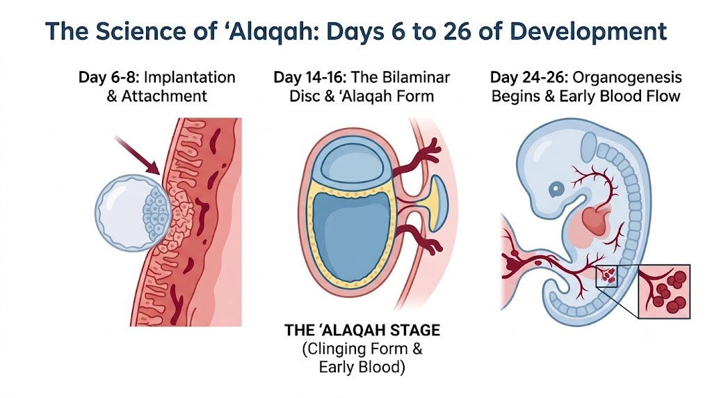
Observe how much the embryo of 24 days (figures 5) looks like a leech (Arabic, alaca, a thing which clings) and that it later appears like a chewed substance (Arabic, mudga, chewed flesh) after most of the somites form during the fourth week (figure 6).
During the embryonic period, the embryo acquires distinctive human characteristics as the bones and muscles begin to form (figure 7).
By the end of the embryonic period, the beginnings of all the main organ systems have been established. The external appearance of the embryo is greatly affected by the formation of the brain, heart, liver, somites, bones, limbs, ears, nose, and eyes. As these structures develop, they affect the appearance of the embryo by characteristics that mark the embryo as unquestionably human. Because the beginnings of all essential external and internal structures are formed during the embryonic period, these five weeks constitute the most critical period of development. Developmental disturbances during this period may give rise to major congenital malformations.
The second major stage of prenatal development is the fetal stage (figure 8). This is a period of rapid growth and differentiation. The eyes are open by 24 weeks, by which time the fetus may be capable of survival if born prematurely.

A human being develops from a single cell, the zygote, which forms when an ovum is fertilized by a sperm. The two verses from the Koran shown in figure 9, and the saying from the Hadith,. make it clear that sperm are derived from a very small part of the fluid or semen that gushed forth or is ejaculated from the penis. They are expelled from the urethra via the same route followed by the urine which is sometimes referred to as a ‘despised’ fluid.
There are several references in the Koran which indicate that a human being develops from a part which is slowly drawn out or extracted (Arabic, sulala). It is reasonable to interpret the nutfa as the small sample of sperms which are extracted from ejaculated semen, because it is well established that only a few hundred of the several million sperms in the semen are able to pass through the uterus and surround the ovum in the uterine tube.
The mixed drop mentioned in the Koran (figure 10) could refer to the mixture of a small quantity of sperms with the oocyte and its associated follicular fluid. There are other references in the Koran to the origin of man from a small quantity of ‘mingled fluids’, undoubtedly the male and female sexual secretions. As we know, a secondary oocyte is expelled from the ovary during a process known as ovulation. The oocyte and the follicular fluid pass into the uterine tube, where, if coitus has occurred, they are mixed with several hundred sperms. The resulting mixture (drop) composed of the ovum and the penetrating sperm, becomes the zygote or primordium of the embryo.
A popular idea in the 17th century among scientists was that the sperm contained a miniature human being that simply enlarged inside the sperm. Another equally strong idea was that the ovum contained a miniature human being that was stimulated to growth by the semen.
It was not until the 18th century that Spallanzani showed experimentally that both male and female sex products were necessary for the initiation of development. From his experiments, including artificial insemination in dogs, he concluded that the sperm was the fertilizing agent. It is difficult not to interpret the mixed drop mentioned in the Koran in the 7th century as a reference to the mingling of the male and female sex cells described eleven centuries later.
The idea that development results from a genetic plan (figure 11) contained in the chromosomes of the zygote was not discovered until the end of the 19th century. The verse from the Koran clearly implies that the nutfa contains then plan or blueprint for the future characteristics and features of the developing human being.
The realization that sex was determined at fertilization (figure 12) was determined about 60 years ago when the sex chromosomes were discovered. The determination of sex is one of the main results in fertilization. The strong male influence of the Y chromosomes on sex development was discovered about a decade ago.
The blastocyst or early embryo implants in the uterus 10 days after fertilization. It assumes a human appearance during the eighth week. Hence, 40 to 45 nights after its implantation in the uterus (figure 13), it would be 50 to 55 days old and have a distinctive human appearance. Prior to this time, the human embryo is similar to the embryos of other mammalian species.
An understanding of the implantation process of the human blastocyst is also implied in the Koran (figure 14). A tilth refers to the cultivation of land and the comparison of implantation of the blastocyst to the planting of a seed is a very appropriate one. Just as soil covers the seed, the uterine epithelium covers the implanted blastocyst (figure 2). The blastocyst soon develops chorionic villi for acquiring nourishment from the maternal blood. Similarly the embryo formed from the seed develops roots for acquiring nourishment from the soil.
It is well established that there is a lag or delay in the development of the embryo during implantation (figure 15). For an entire week, very few changes can be observed in the developing embryo or bilaminar embryonic disc. The agreement between the lag or gap in development mentioned in the Koran and the slow rate of change occurring during the second and third weeks is amazing. These details of human development were not described until about 40 years ago.

Another verse in the Koran (figure 16) refers to the leech-like appearance (figure 5) and the chewed-like stages of human development (figure 6). Another one (figure 17) states that during this chewed stage the embryo has both differentiated and undifferentiated parts. It is well established that the brain and the heart are only partially differentiated at the end of the fourth week when the embryo resembles chewed substance (figure 6).
After the chewed-like appearance, bones develop which are soon clothed with muscles (figure 18). The bones begin to form in the sixth week and muscles attach to them shortly after this. By the beginning of the seventh week, the bones give a human shape to the embryo’s body (figure 7).

The ears and eyes being to form in the fourth week and are clearly visible at six weeks, 42 days after the zygote or nutfa forms (figure 19). Sex is not distinguishable at this age. Is this the basis of the angel’s questions?

Human features become recognizable about 42 days after the zygote forms (figure 20), but are not distinctly human until the eighth week. Note that the sex of the embryo is not clearly distinguishable at this stage. The external genitalia are not distinctly male or female until the 12th week.
There are other statements in the Koran and sayings in the Hadith about embryology that are meaningless to me, but very likely they will make sense later when new knowledge is developed. The agreement I have found between the statements in the Koran and sayings in the Hadith may help to close the gap between science and religion which has existed for so many years.
REFERENCES
AREY, L.B., Developmental Anatomy: A Textbook and Laboratory Manual of Embryology, revised 7th Philadelphia, W.B. Saunders Co., 1974.
MEYER, A.W., The Rise of Embryology, Stanford, Stanford University Press, 1939.
MOORE, K.L., The Developing Human. Clinically Oriented Embryology, 3rd edn, Philadelphia, W.B. Saunders Co., 1982.
NEEDHAM, J., A History of Embryology, 2nd edn, Cambridge, Cambridge University Press, 1959.
NISHIMURA, H., TANIMURA, T., SEMBA, R. AND UWABE., Normal development of early human embryos: Observation of 90 specimens at Carnegie Stages 7 to 13, Teratology, 10, 1974.
OPPENHEIMER, J. M., Problems, concepts and their history. In B.H. WILLIER, P.A. WIESS AND V. HAMBURGER (eds), Analysis of Development, Philadelphia, W.B. Saunders Co., 1965.
O’RAHILLY, R., Guide to the staging of human embryos, Anat. Anz., 130, 556, 1972.
O’RAHILLY, R., Developmental Stages in Human Embryos. Part A: Embryos of the First Three Weeks (Stages 1 to 9), Washington, D.C., Carnegie Institute.
STREETER, G.L., Developmental horizons in human embryos: Description of age group XI, 13 to 20 somites, and age group XII, 21 to 29 somites, Contrib. Embryol. Carnegie Inst., 30.211, 1942.


What do you think?